

Customer Support
Hotline

Send message via zalo

Description
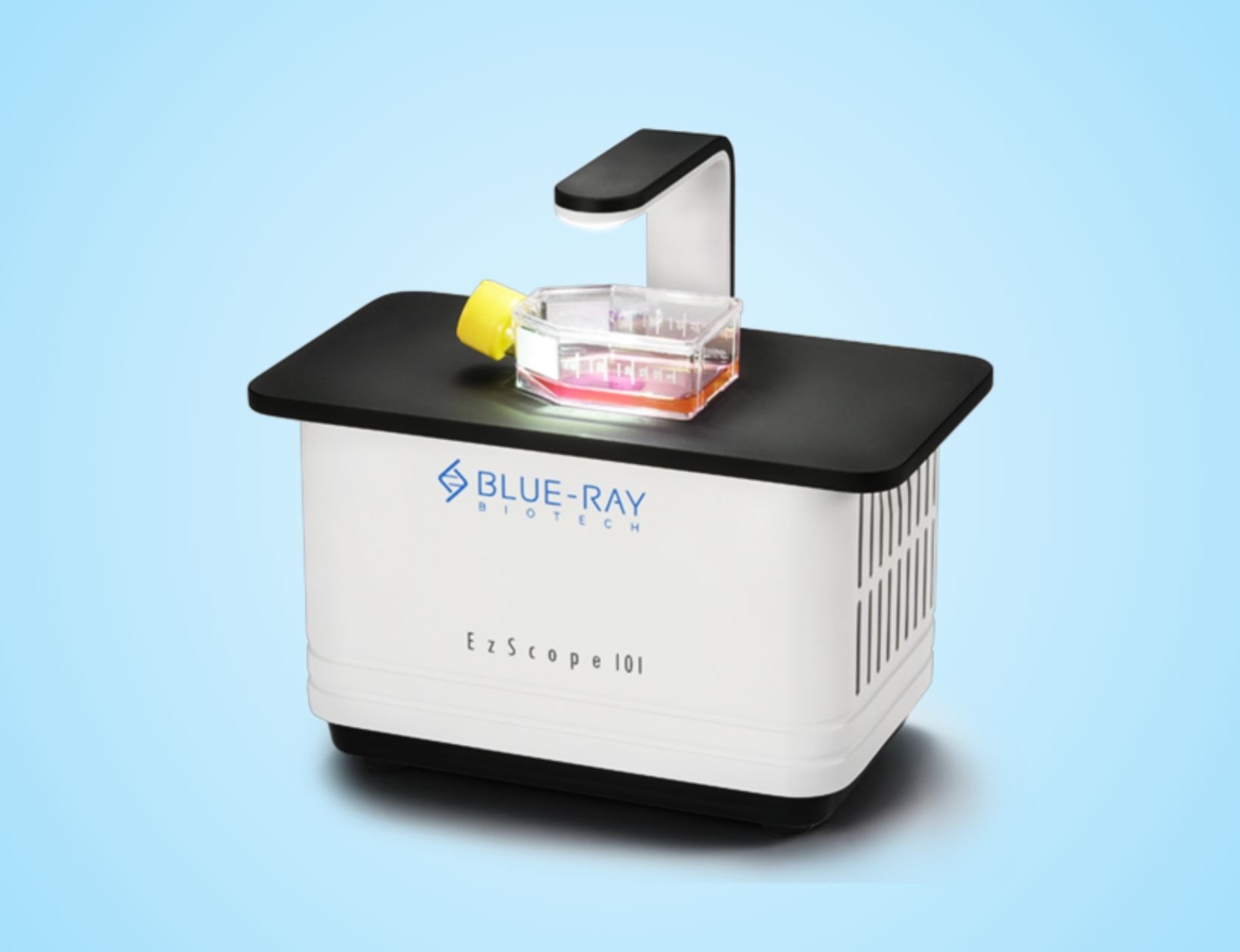
EzScope 101 – Live cell imaging system Live cell live show – EzScope 101 is a dedicated live cell imaging system that helps to streamline your research workflow with improved efficiency and productivity, no more hassles to remove cells from incubator for observation. EzScope 101 brings 24/7 measurements under precisely controlled conditions in a non-perturbing environment. You can observe the images anytime with walk-away convenience. Up to four samples can be monitoring simultaneously in a same incubator.
Features and benefits
Incubator Live View Designed to be used inside the incubator, without the need to remove your cells from incubator to enhance culture quality control. Minimizes Experimental Variations Up to four units of EzScope can to be setup in the same incubator and controlled by one computer. This enables the monitoring of samples simultaneously, reduces errors caused by environment variations. Exceptional Image Quality Adopts high contract brightfield optical configuration, coupled with precise motorized focusing, and two interchangeable magnifying objective lenses. Remote Monitoring of Experiment Allows flexible remote monitoring the assay via Windows-based remote desktop software. Easy Image Editor Captures and edits images easily with EzCapture software: – Live preview for up to 4 units of EzScope – Capture single image or time-lapse series – Flatfielding correction for even brightfield background – Time-lapse video output – Spatial calibration – Measure and convergence analysis
Applications
EzScope 101 enables microscope-in-incubator to live view for real-time cell monitoring, migration, growth, and invasion, plus a wide range of phenotypic cell-based assays. https://youtu.be/xCDo1PlQ2OI Sample: Human Ovarian Cancer Cell (HM-4) Description: The time-lapse video shows the formation of a 3D spheroid by using a self-made suspended tumorspheroid chip. Similar Applications: Cell migration, wound healing, cell confluence studies https://youtu.be/aaBWfukuw9E Sample: Cardiac Muscle cell Description: The time-lapse video clip of differentiated cardiac muscle stem cells reveals a beating feature. Similar Applications: All kind of stem cell related research https://youtu.be/mHuIPfxXREo Sample: Human Embryonic Lung Organoids (Control and IGFBP3 siRNA Transfection) Description: This time-lapse video demonstrates the differentiation of human embryonic lung organoids after transfection with IGFBP3 siRNA. The EzScope 101 Live Cell Imaging System continuously captured morphological changes over 48 hours, allowing detailed observation of the transfection effects without disturbing the incubator environment. Similar Applications: Organoid differentiation, cell growth studies, real-time live-cell imaging https://youtu.be/qihtSFJMr6c Sample: Lung Tumor Organoids Description: The time-lapse video captures the growth and morphological changes of lung tumor organoids using the EzScope 101 Live Cell Imaging System. Continuous imaging inside the incubator enables real-time monitoring of organoids development, providing valuable insights into cancer cell behavior. Similar Applications: Cell growth studies, cancer drug screening, organoid-based disease modeling
Additional information
| Status | New |
|---|---|
| Optics | Brightfield (transmitted) with white LED |
| Objective Lens | 10x, 20x (optional) |
| Camera | 1.3 MP CMOS Sensor |
| Image Resolution | 1280 x 1024 pixels |
| Export Formats | Tiff (image), AVI (video) |
| Software | EzCapture with snapshot, time-lapse and confluence, etc |
| Field of View | 2.6 x 2.0 mm (10x objective) |
| Resolution | 2 µm/pixel (10x objective), 1 µm/pixel (20x objective) |
| Live View Frame Rate |
Related products

Customer Support
Hotline
Send message via zalo